
Cervical Spine Simulator
Mixed Reality-based training device with tangible haptic feedback through actuation, for physiotherapy students to practice the mobilisation techniques in the cervical region.
Technologies Used
Project Overview
The Cervical Spine Simulator is an innovative HCI research project that combines mixed reality with tangible haptic devices to create an immersive physiotherapy training system. This project addresses critical gaps in physiotherapy education by providing hands-on experience with cervical spine actuation device through a safe, repeatable, and realistic training environment.
Built using Unity VR and air pressure based actuation systems, the simulator enables physiotherapy students and professionals to practice cervical spine mobilsation techniques with real-time haptic feedback. The system adds to the traditional classroom learning and hands-on physiotherapy experience, offering a risk-free environment for skill development.
This research contributes to the field of HCI by exploring how tangible interfaces using air pressure actustion can enhance immersive virtual environments, particularly in different training scenarios. The project demonstrates the potential of combining MR visualization with physical haptic feedback to create more effective and engaging training experiences.
Problem & Motivation
Physiotherapy students often rely on peers to learn manual mobilisation techniques in the cervical region of the neck. This dependency creates an inconsistent learning experience, restricting students from safely exploring variations in technique.
There is a need for a controlled training environment that allows students to independently practice cervical manipulations to build confidence through safe and repeatable experiences.
Research Methodology
Research Methodology
Study Design
A mixed-method design moved from domain research into fieldwork, prototype development, and evaluation.
Domain Research
Reviewed cervical examination techniques and mixed-reality physiotherapy training precedents.
Semi-structured Interviews
Worked with a physiotherapy lecturer to understand mobilisation techniques and teaching constraints.
Fieldwork
Observed class practice and ran contextual interviews to locate curriculum fit.
Prototype
Built physical haptic and MR interface prototypes in parallel.
Evaluation
Tested the physical device and mixed-reality interface through task-based scenarios.
Discover
In the Discover phase of our research, we conducted primary research into the domain of physiotherapy to get an understanding of existing learning methods and challenges. This phase also included exploring novel methods of interaction across different technologies to identify opportunities for enhancing physiotherapy education through innovative and immersive experiences.
Primary Research
• For the domain of physiotherapy our research was mainly sourced from online resources like google and YouTube to understand different techniques in the cervical region. The main resource we followed to familiarise ourselves was Physiopedia. https://www.physio-pedia.com/Cervical_Examination
• To explore new and novel methods of interaction we explored the different applications of Mixed Reality in physiotherapy education.
Discovery
Initial Assumptions
Practice Needs Repetition
Students need a safe way to repeat cervical mobilisation techniques outside peer practice.
Physical Feedback Matters
A tangible device can make stiffness and bone response easier to understand than visual simulation alone.
MR Can Add Context
Mixed reality can show internal movement while keeping the learner's hands on the training object.
Interview goals
Understand the physiotherapy techniques in the cervical region.
Gain insights into teaching and learning challenge.
Visualise the physical impact of different techniques.
Semi Structured Interviews
• We conducted semi structured interviews with physiotherapy lecturer to understand how the techniques are performed gather a deeper insight into it.
• These interviews were conducted in the interaction lab.
• To assist our understanding we made used a 3D printed model of the bone in the cervical region, allowing the physiotherapist lecturer show us the exact impact of different techniques.

Bones 3D printed using PLA tough plastic and combined using glue and foam.

Bones 3D printed using PLA tough plastic and combined using glue and foam.
Key Findings from the Interviews.
Focus on techniques in the prone position.
The physiotherapy lecturer suggested to focus on techniques in the prone position as it is the most common position for cervical spine mobilisation.
Focus on Palpation technique from Maitland's Vertebral Mobilisation.
We were suggested to focus on Palpation technique from Maitland's Vertebral Mobilisation as it is a common technique used in physiotherapy.
Focus specifically on Central PA and Unilateral PA.
Two types of force application techniques were suggested to be focused on: Central PA and Unilateral PA.
Initial Sketches of the System
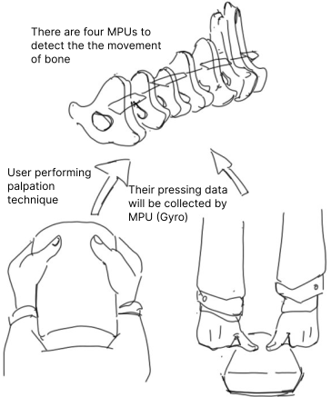
Realtime Movement of the bones
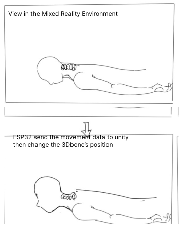
Realtime Movement of the bones
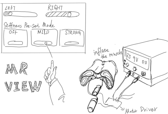
Stiffness Level Adjustment using MR Interface
Fieldwork for Design
Our fieldwork consisted of Observational Study and Contextual Interviews.
The observational study was conducted to understand the practice of various physiotherapy techniques that are included in the student's curriculum. Contextual interviews were conducted with the physiotherapy lecturer to gain insgihts into how these techniques are taught to the students.
Observational Study
We were able to attend one of the classes of physiotherapy. Due to ethical considerations we were not allowed to record any video or audio from the observations. The study took place over a time of 30 mins at the Department of Physiotherapy.
Contextual Interviews
These interviews were conducted to understand how this system would fit in curriculum for the students.
Findings from the fieldwork
Fieldwork
Observations and Interviews
Observational Study
Observed physiotherapy class practice for 30 minutes under ethical recording constraints.
Contextual Interviews
Discussed how the simulator could fit into physiotherapy teaching with a lecturer.
Design Direction
Focused on prone-position cervical mobilisation, palpation, and Central PA / Unilateral PA techniques.
Iterative Prototype
Prototyping was divided into two stages : Physical and MR Prototyping.
Physical Prototype
The physical prototype included designinf the structure of the muscles, case to hold the bones and also making the silicon skin to cover the muscles and simulate the skin texture.
First Iteration
Physical Prototype
First Iteration
Muscle Structure
Designed the internal muscle structure around the cervical bone model.
Bone Housing
Built a case to hold the vertebrae model and support hand pressure.
Silicone Skin
Added a silicone skin layer to approximate touch and surface texture.
Feedback on the First Prototype
Prototype Feedback
Feedback on the First Prototype
Uneven Stiffness
The silicone skin did not fit perfectly, creating inconsistent stiffness feedback.
Bone Detectability
Protruding bones were easy to feel, while recessed bones were harder to identify.
Loose Base
The lower seat moved when air entered the material, reducing stiffness accuracy.
We consulted with a physiotherapy lecturer about the prototype. These were the feedback recieved
Second Iteration
Physical Prototype
Second Iteration
Modular Chambers
Segmented the muscle structure so air pockets could be moved between chambers.
Replaceable Air Pockets
Designed inlets for 3D-printed air pockets that hold and release pressure.
Localized Stiffness
Allowed different stiffness locations to be tuned externally with driver motors.
We had consultations with the physiotherapist lecturer to improve the previous iteration. In this iteration we designed a modular system for the muscles. This modular design offers the following:
1. Inlets for 3D printed air pockets to hold on to air. 2. Ability to switch the air pockets in different chambers in the model.
Implications: This design caters to different stiffness locations in the neck. The air pushed into chambers can be modified using driver motors externally. Depending upon the feedback from the physiotherapy lecturer the stiffness can also be felt after pushing a bit on the meta material due to its modular design.
Mixed Reality Prototype
Our MR Prototype consist of two parts, one is the movement of the bones and the second is an interface to control the motors from the mixed reality interface.
We used Unity to develop our system and ShapesXR to prototype the interface design.
Bone Movement in Unity
Unity Prototype
Bone Movement in Unity
Input
Physical pressure and technique selection drive the simulation state.
Visualization
Unity shows cervical bone movement to make internal response visible.
Feedback Loop
Learners compare what they feel in the device with what they see in MR.
ShapeXR Interface Prototype
MR Interface
ShapeXR Interface Prototype
Technique Controls
Early MR panels explored choosing mobilisation techniques and target regions.
Stiffness Adjustment
Interface controls mapped stiffness levels to the physical actuation system.
Session Feedback
Prototype screens made the learner's selected task and current system state visible.
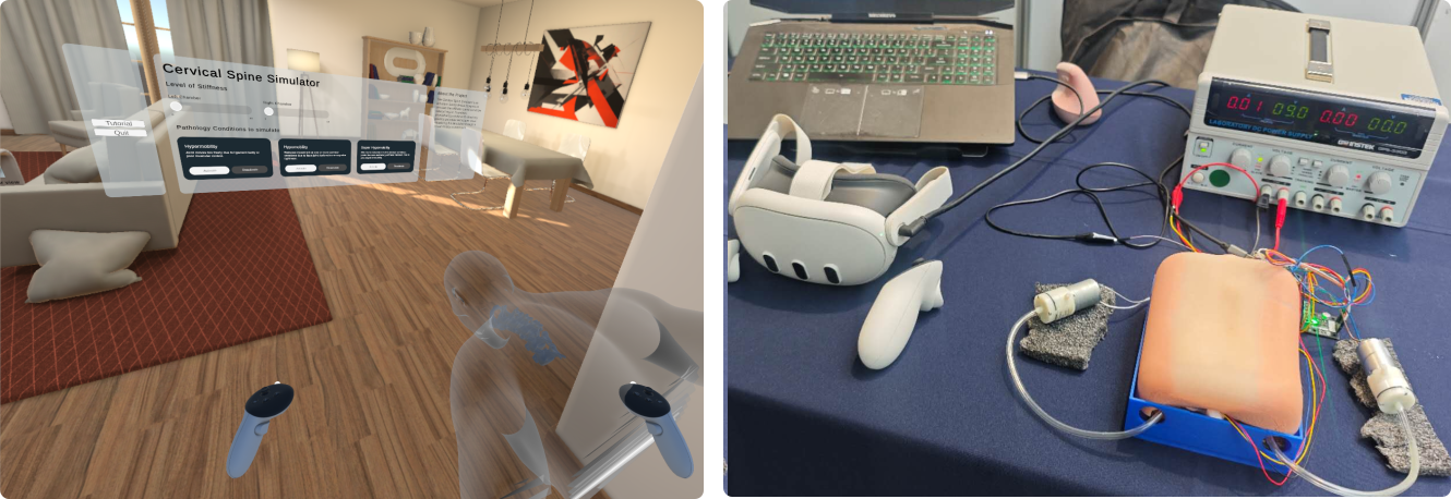
Unity Mixed Reality Interface
MR Interface
Unity Mixed Reality Interface
Select Region
Learners choose the relevant cervical region inside the mixed-reality scene.
Tune Stiffness
Controls send stiffness changes to the actuation hardware.
Inspect Motion
The MR view shows bone response while practice continues on the physical device.
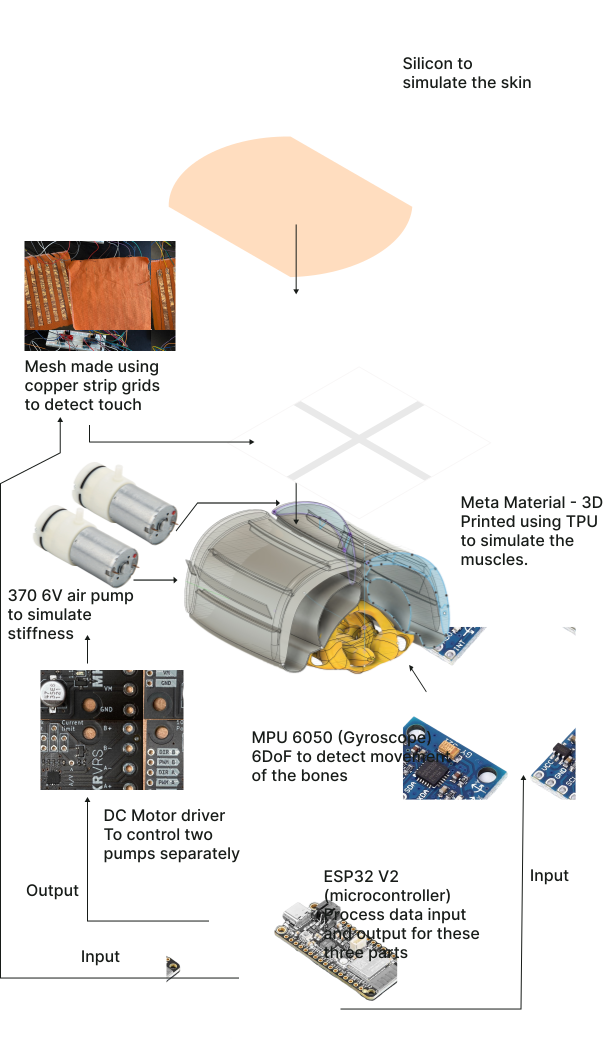
System Design
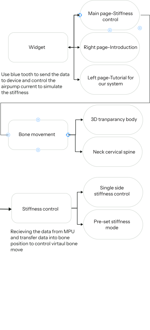
Final Prototype
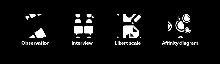
Evaluation Methods
Participant Recruitment
We recruited 9 participants, mostly students of IT and a physiotherapy lecturer, due to ethical limitations we were not able to recruit physiotherapist students.
Evaluation Study Design
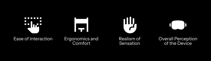
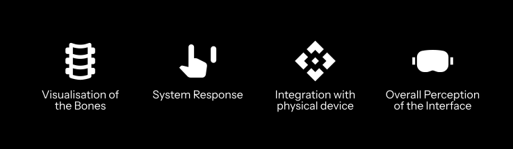
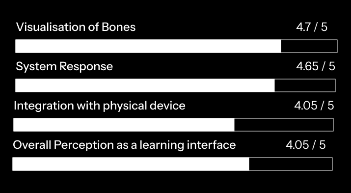
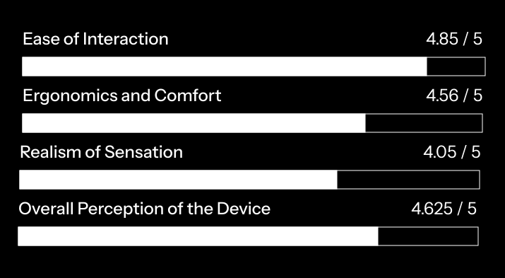
We conducted the study with both the physical device and Mixed Reality Environment to understand the credibility of the system working in unision. Task based scenarios were designed, providing Likert Scale Templates for the physical device and the Mixed Reality environment individually. Following are the domains of were decided to test for the physical device and the Mixed Reality Interface
Evaluation Likert Scale Results
Findings from the Interviews and Observations
For Physical Device
• The silicon skin does not fit perfectly on the device, thus giving uneven feeling of stiffness. • The bones which protrude the meta-material are easy to feel but the others are a bit hard to feel. • The bottom seat of the device is bit loose when the air pumped into the meta material, thus giving less accurate stiffness simulation.
For MR Interface
• Include a tutorial on using the MR Interface, as it would be hard for a first time user to find their way around. • The visualisation of the movement of the bones is not as accurate to the movement of real bones. • More elements in the MR Interface could be made grababble.